论文标题:Radiological findings in ancient Egyptian canopic jars: comparing three standard clinical imaging modalities (x-rays, CT and MRI)
期刊:European Radiology Experimental
作者:Patrick E. Eppenberger et al
发表时间: 2018/6/20
数字识别码:10.1186/s41747-018-0048-3
原文链接:https://eurradiolexp.springeropen.com/articles/10.1186/s41747-018-0048-3?utm_source=Other_website&utm_medium=Website_links&utm_content=DaiDen-BMC-European_Radiology_Experimental-Other_Biology-China&utm_campaign=AJH_USG_BSCN_DD_eurradiolexp_Egyptian
研究古埃及卡诺匹斯罐中的内容物对埃及古物学和生物医学研究都有价值,但打开罐子可能会破坏其中宝贵的生物材料。在European Radiology Experimental杂志发表的一篇新文章中,研究人员使用医学成像技术来探查这些古老的容器。

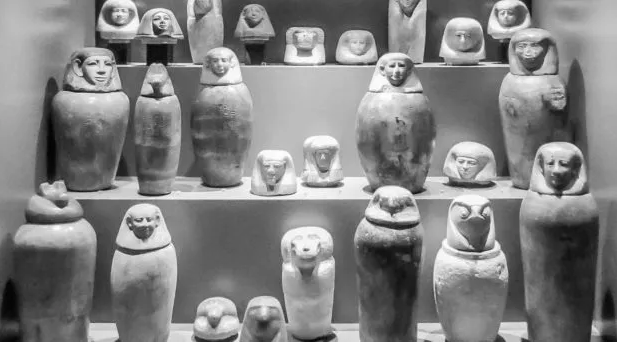
克罗地亚萨格勒布埃及考古博物馆中展出的古埃及卡诺匹斯罐
图片来自论文
“卡诺匹斯罐项目”由瑞士国家科学基金会资助,是世界上首次在真正的跨学科研究环境中,对从欧洲和美国博物馆收集的大量古埃及的卡诺匹斯罐进行研究。
这项关注卡诺匹斯罐内容物的研究获得了传统古代木乃伊研究方法无法得到的结果。该项目在预先进行埃及古物学评估的基础上,对古埃及的罐子和木乃伊进行了宏观、放射、化学和古生物学研究。
古埃及的卡诺匹斯罐
古埃及人对死者的尸体进行防腐处理,因为他们相信灵魂游荡于肉体之外,一定要能够返回肉身。因此,尸体死后的保存对于灵魂在死后的存在至关重要。另一方面,为了避免内脏的腐烂,必须从身体中取出内脏并且同样需要保存。
死者的某些内脏被保存在一种叫做卡诺匹斯罐的容器中。虽然古埃及丧葬实践和卡诺匹斯罐的设计和使用从最初的实验阶段(公元前2700-2200年)到其使用高峰(公元前1550-1077年)以及第三中间期(公元前1077-652年)发生了巨大改变,但通常会用到四个卡诺匹斯罐,每个罐子专用于保管一个特定器官。
大多数罐子都是用雪花石膏或陶土制成的,高30-40厘米,罐子的盖子分为四种类型,表明了它们的内容:人头代表肝脏,狒狒代表肺,豺狼代表胃,而猎鹰代表肠子。卡诺匹斯罐被放置于墓室中的石棺旁。
法国语言学家Jean-Francois Champollion(1790-1832)破译了罗塞塔石碑上的象形文字,他似乎早在1812年就发现了这些文字的用途,但对这些文字内容的研究最近才开始的,而迄今为止,几乎没有人分析过卡诺匹斯罐。长期以来,对它们的研究仅限于艺术角度。

卡诺匹斯罐三维表面重建和容积计算
图片来自论文
进化医学研究所所长、这项研究的主要作者Frank Ruhli教授表示:“令人惊讶的是,迄今为止在生物医学研究中,古埃及含有珍贵的木乃伊人体内部器官神奇罐子常常被忽视。尽管它们有其独特的价值,能够帮助人们理解疾病正在发生的演化过程。”
研究古埃及卡诺匹斯罐的好处在于,它在一定程度上使科学家们摆脱了对古埃及木乃伊进行侵入性研究的伦理约束,从而开辟了几个有吸引力的探索领域。
医学领域将受益于对病原体演化的深入了解,而遗传指纹和病原体鉴定对增进我们对古埃及健康和社会结构的理解至关重要。
最重要的是罐子的内容物
然而,适合于这类研究卡诺匹斯罐及其内容物是有限的。打开罐子可能导致其中的生物组织被氧化,甚至细菌污染。为了避免浪费这种独特的研究材料,第一步是使用最新的医学成像技术来查看罐子内部,即平面X射线、计算机断层扫描(CT)和磁共振成像(MRI)。
该项研究首次比较了这三种标准临床影像学方法在古埃及卡诺匹斯罐内容物研究方面的应用,并探讨了适用于这些特殊样品的、古放射性学中三种主要先进诊断方法的一般可行性和诊断敏感性。
出乎意料的是,放射分析带来了社会文化相关发现:与代表了古埃及木乃伊化过程中最古老信息的希罗杜斯的记载相反,罐子中保存的可能不是完整的器官,而仅仅是一些器官碎片。即使在干燥后,大多数卡诺匹斯罐的体积都无法容纳完整的人类器官。这一发现具有重大意义,埃及人在死后要寻找的不是器官本身,而仅仅是一种象征意义,并非真实存在的器官。这可能意味着,对于死亡和来世的理解与之前所认为的处于不同的抽象水平,然而这一点仍有待证实。
摘要:
Background
The aim of our study was to evaluate the potential and the limitations of standard clinical imaging modalities for the examination of ancient Egyptian canopic jars and the mummified visceral organs (putatively) contained within them.
Methods
A series of four ancient Egyptian canopic jars was imaged comparing the three standard clinical imaging modalities: x-rays, computed tomography (CT) and magnetic resonance imaging (MRI). Additionally, imaging-data-based volumetric calculations were performed for quantitative assessment of the jar contents.
Results
The image contrast of the x-ray images was limited by the thickness and high density of the calcite mineral constituting the examined jars. CT scans showed few artefacts and revealed hyperdense structures of organ-specific morphology, surrounded by a hypodense homogeneous material. The image quality of MRI scans was limited by the low amount of water present in the desiccated jar contents. Nevertheless, areas of pronounced signal intensity coincided well with hyperdense structures previously identified on CT scans. CT-based volumetric calculations revealed holding capacities of the jars of 626–1319 cm3 and content volumes of 206–1035 cm3.
Conclusions
CT is the modality of choice for non-invasive examination of ancient Egyptian canopic jars. However, despite its limitations, x-ray imaging will often remain the only practicable method for on-site investigations. Overall, the presented radiological findings are more compatible with contained small organ fragments rather than entire mummified organs, as originally expected, with consequent implications for envisioned future sampling for chemical and genetic analysis.
阅读论文原文,请访问
https://eurradiolexp.springeropen.com/articles/10.1186/s41747-018-0048-3?utm_source=Other_website&utm_medium=Website_links&utm_content=DaiDen-BMC-European_Radiology_Experimental-Other_Biology-China&utm_campaign=AJH_USG_BSCN_DD_eurradiolexp_Egyptian
(来源:科学网)
特别声明:本文转载仅仅是出于传播信息的需要,并不意味着代表本网站观点或证实其内容的真实性;如其他媒体、网站或个人从本网站转载使用,须保留本网站注明的“来源”,并自负版权等法律责任;作者如果不希望被转载或者联系转载稿费等事宜,请与我们接洽。