|
|
|
|
|
AHM | 军事医学研究院车津晶教授团队:低温制备负载胸腺素 β4 的可溶性微针特异性结合下调的免疫调节因子 Vsig4 和 IL22rα2 促进伤口愈合 |
|
|


研究背景:
皮肤伤口愈合是临床常见难题,传统治疗面临大分子药物透皮难、肽类药物稳定性差、免疫微环境调控不足等挑战。胸腺肽β4(Tβ4)虽具有抗炎、促血管生成、抑制凋亡等多重修复功能,但其临床应用受限于递送效率低和作用机制不明。
文章概述:
军事医学研究院车津晶团队报道了一种基于低温干燥工艺的Tβ4可溶性微针系统,并首次揭示Tβ4通过特异性结合并下调免疫调控因子Vsig4和IL22rα2,重塑伤口免疫微环境、协同促进组织修复的全新作用机制。该研究为难愈性创面的治疗提供了“高效递送+精准免疫调控”的创新策略。
图文导读:
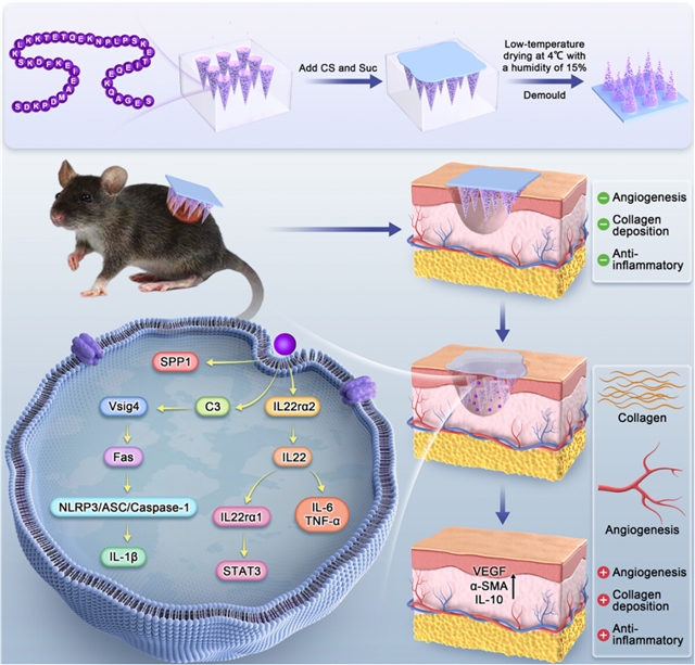
FIGURE1:Schematic diagram depicting the fabrication and application of thymosin β4 (Tβ4)-loaded microneedle patches for wound treatment. The Tβ4 was loaded into chitosan (CS) and sucrose MNs under mild conditions (4°C, 65% relative humidity). The Tβ4 MN patch specifically binds to the downregulated immune regulators Vsig4 and IL22rα2, thereby accelerating wound healing.
研究采用4℃、65%相对湿度的温和条件,以壳聚糖和蔗糖为基质,成功制备出负载Tβ4的可溶性微针(图2)。该工艺有效避免了传统微针制备高温或紫外固化对多肽活性的破坏。微针形态均匀、针尖尖锐,具备良好的机械性能,可轻松穿透皮肤角质层,并在伤口部位快速溶解、释放药物(图3、图4)。
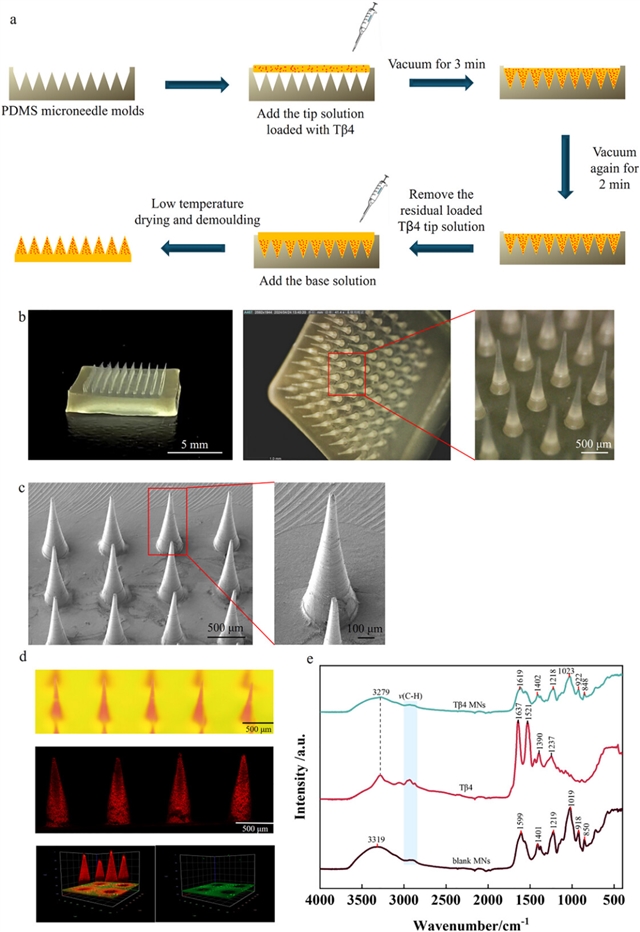
FIGURE2:Preparation and morphological characterization of Tβ4 microneedles, drug distribution, and material interactions. (a) Schematic illustration of the preparation process for our Tβ4 MN patch. (b) Optical microscopy image of a fabricated Tβ4 MN patch. (c) SEM image of a fabricated Tβ4 MN patch. (d) Fluorescence microscopy image of the distributions of doxorubicin hydrochloride (red, representing Tβ4) and FITC (green, representing the MN base) in the patch MNs. (e) FTIR spectra of Tβ4, Tβ4 MN, and blank MN samples.
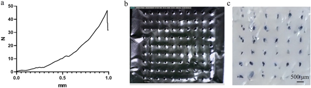
FIGURE3:Testing of the mechanical properties and hardness of our fabricated Tβ4 MNs. (a) Force-displacement curves of the MNs. (b) Penetration of the MNs into the aluminum foil. (c) Trypan Blue staining of the MN insertion wounds in ex vivo porcine skin.
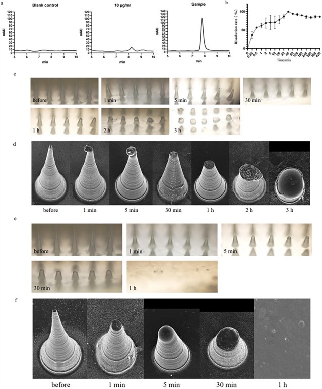
FIGURE4:MN drug loading, in vitro release, and in vivo degradation experiments. (a) Representative chromatogram of the Tβ4 MNs. (b) In vitro drug release profile of the Tβ4 MNs (n = 3). (c) Morphology of the Tβ4 MNs as observed via optical microscopy before and after insertion into murine skin in vivo. (d) Morphology of Tβ4 MNs observed via SEM before and after insertion into murine skin in vivo. (e) Morphology of the Tβ4 MNs before and after insertion into a murine wound model, as observed via optical microscopy. (f) Morphology of the Tβ4 MNs before and after insertion into a murine wound model, as observed via SEM.
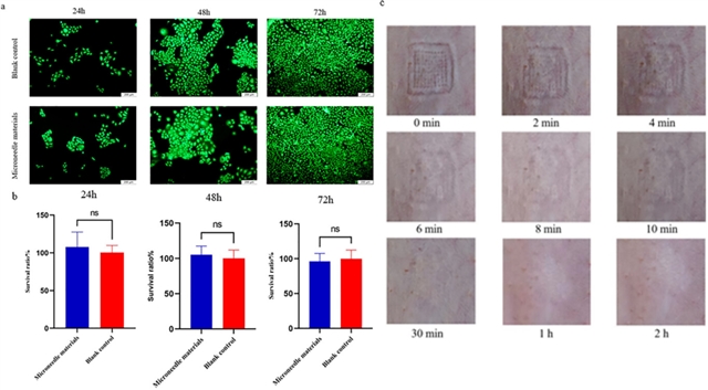
FIGURE5:Biocompatibility of Tβ4 MNs. (a) Survival staining ratio of HaCAT cells after different treatments. (b) Cell viability was determined using CCK8 assays. ns, no statistical significance. (c) Skin recovery within 2 h following puncture by the MNs. Data were represented as mean ± standard deviation and analyzed using an independent samples t-test. Statistical significance is indicated by ns, no statistical significance *p < 0.05, **p < 0.01, and ***p < 0.001, n = 3.
在小鼠全层皮肤缺损模型中,Tβ4微针展现出显著的促愈合效果。治疗第10天,微针给药组的伤口闭合率达95.71%,显著优于Tβ4溶液组和阳性对照药重组人III型胶原蛋白组(图6)。组织学分析进一步证实,微针治疗能有效缩小创面间隙、促进胶原沉积,并上调VEGF、α-SMA、IL-10等关键修复因子的表达(图7、图8)。
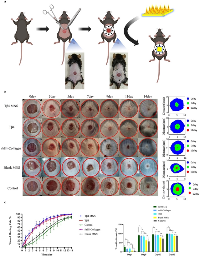
FIGURE6:Effect of Tβ4 MN patch on wound healing in a murine model. (a) Schematic diagram of the wound model and treatment strategy. (b) Gross view of wounds in mice treated using our Tβ4 MN patch, topical Tβ4 only, blank MN patch, topical rhIII-collagen, or control at different time points. (c) Measurement of the wound areas shown in panel b. Data were represented as mean ± standard deviation and analyzed using a one-way analysis of variance (ANOVA) with Tukeys post hoc test. Statistical significance is indicated by ns, no statistical significance *p < 0.05, **p < 0.01, and ***p < 0.001, n = 8.
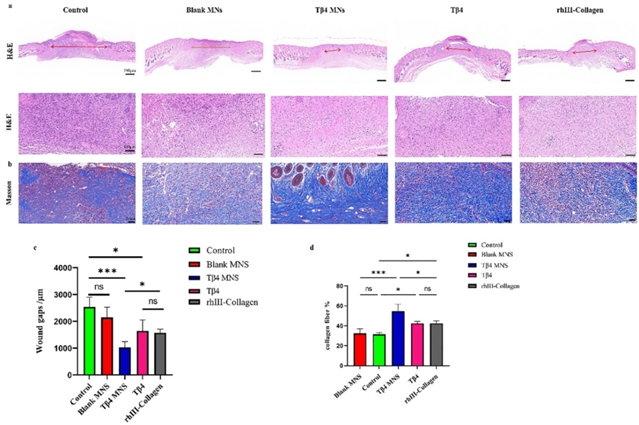
FIGURE7:Wound-healing as assessed through HE and Massons trichrome staining. (a) HE staining of murine wounded skin on day 14 post-treatment (single-headed arrows indicate un-epithelialized areas). (b) Massons trichrome staining of murine wounded skin on day 14 post-treatment. (c) Assessments of wound gaps in the control, blank MN, Tβ4 MN, Tβ4 only, and rhIII-collagen groups on day 14 post-treatment. (d) Assessment of collagen deposition via Masson staining at day 14 post-treatment among the control, blank MN, Tβ4 MN, Tβ4 only, and rhIII-collagen groups. Data were represented as mean ± standard deviation and analyzed using a one-way analysis of variance (ANOVA) with Tukeys post hoc test. Statistical significance is indicated by ns, no statistical significance *p < 0.05, **p < 0.01, and ***p < 0.001, n = 3.
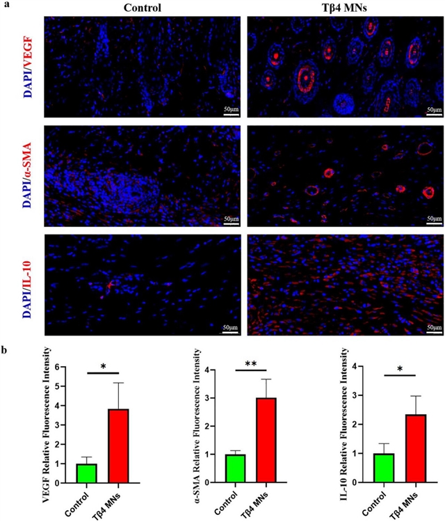
FIGURE8:Immunofluorescent staining of wound sections at day 14 post-treatment. (a) Fluorescence intensity for VEGF, α-SMA, and IL-10 expression in skin tissues at the wounded sites on day 14 in the control and Tβ4 MN groups. (b) Fluorescence intensity analysis for VEGF, α-SMA, and IL-10 in the control and Tβ4 MN groups. Data were represented as mean ± standard deviation and analyzed using an independent samples t-test. Statistical significance is indicated by *p < 0.05, **p < 0.01, n = 3.
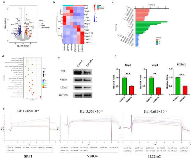
FIGURE2:Principal component analysis, heatmap, volcano plot of differentially expressed mRNAs, and our analysis of known genes related to wound healing of skin tissues in the control and Tβ4 MN groups. (a) Volcano plot of the DEGs regulated by Tβ4 MNs, filtered by |log2F − C| > 2, and p < 0.05. (b) Heatmap of DEGs related to wound healing in skin tissues. (c) Enriched GO terms related to the identified DEGs. (d) Enriched KEGG pathways related to the identified DEGs. (e) WB results for skin tissue proteins. (f) Real-time quantitative plot of the sequential expression of the identified DEGs. (g) Results of surface plasmon resonance (SPR) analysis to investigate the protein binding characteristics of Tβ4. Data were represented as mean ± standard deviation and analyzed using an independent samples t-test. Statistical significance is indicated by *p < 0.05, **p < 0.01, and ***p < 0.001, n = 3.
基于上述发现,研究提出了全新的“双轴协同调控”模型:一方面,Tβ4通过下调Vsig4,可能抑制NLRP3炎症小体过度活化,减轻炎症损伤;另一方面,通过下调IL-22的“诱骗受体”IL22rα2,增强IL-22/STAT3信号通路活性,从而促进上皮增殖与抗菌修复。这一发现突破了以往对Tβ4作用机制集中于血管生成的认知,将其功能提升至免疫微环境精准调控的新层面。
结论:
该研究不仅开发了一种稳定、高效、可临床转化的肽类药物经皮给药递送平台,更重要的是为伤口愈合的免疫调控机制提供了新见解,为开发新一代免疫调节型创面治疗产品奠定了坚实基础。
军事医学研究院毒物药物研究所车津晶研究员和沈阳药科大学张怡轩教授为该论文的共同通讯作者,赫世龙硕士研究生为本论文的第一作者,原梅、冯昌康为第二、第三作者。
论文信息
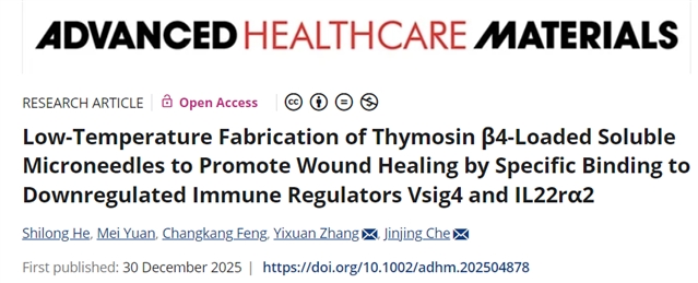
Low-Temperature Fabrication of Thymosin β4-Loaded Soluble Microneedles toPromote Wound Healing by Specific Binding to Downregulated Immune Regulators Vsig4 and IL22rɑ2
Shilong He, Mei Yuan, Changkang Feng, Yixuan Zhang, Jinjing Che
Advanced Healthcare Materials
DOI:10.1002/adhm.202504878
原文链接
https://doi.org/10.1002/adhm.202504878
期刊简介
Advanced Healthcare Materials是 Wiley 旗下 Advanced 系列核心期刊,聚焦医疗健康领域的高影响力材料、设备与技术,是生物材料、纳米医学、组织工程等跨学科领域的权威交流平台。该刊被 SCIE 数据库收录,刊载内容涵盖生物材料、纳米医学、医疗设备等方向,兼顾基础研究与临床转化,对国内学者友好,是领域内成果传播的重要载体。

特别声明:本文转载仅仅是出于传播信息的需要,并不意味着代表本网站观点或证实其内容的真实性;如其他媒体、网站或个人从本网站转载使用,须保留本网站注明的“来源”,并自负版权等法律责任;作者如果不希望被转载或者联系转载稿费等事宜,请与我们接洽。