
近日,比利时根特大学Ruslan I. Dmitriev团队研究了荧光寿命成像显微镜(FLIM)观察纳米塑料在活体肠道类器官中的内化和生物学影响。相关论文于2025年8月12日发表在《光:科学与应用》杂志上。
微纳米塑料(MNP)污染的增加带来了重大的健康风险,但它们的积累机制和对吸收组织的影响仍然知之甚少。解决这一知识差距需要易于处理的模型与动态活细胞成像方法相结合,从而实现多参数单细胞分析。
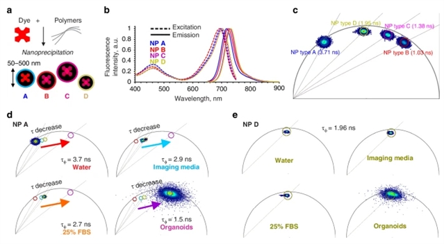
研究组报道了一种将成体干细胞衍生的小肠类器官培养与活荧光寿命成像显微镜(FLIM)相结合的新方法来研究MNP与肠道上皮的相互作用。为了促进这一点,他们优化了猪和猪主题小肠类器官的“尖向外”拓扑结构的实时成像。随后,研究组制作了一组基于PMMA和PS (<200Nm,掺杂深红色荧光染料),并评估它们与显示受控上皮极性的类器官的相互作用。结果发现纳米颗粒与类器官的顶膜和基膜的相互作用不同,并显示出物种特异性的细胞摄取模式。使用相量分析方法证明了FLIM的灵敏度优于传统的基于强度的显微镜。
由此产生的“荧光寿命条形码”能够区分不同类型的MNP及其在类器官中的相互作用位点。最后,研究组分析了短期(1天)和长期(3天)暴露PMMA和基于PS的MNPs对线粒体功能、细胞总能量收支和上皮炎症的影响。他们发现即使是原始的MNPs也会破坏肠上皮细胞的趋化因子产生和线粒体膜电位。所提出的FLIM方法将推进MNP毒性及其对胃肠道组织的生物学影响的研究,并使在活体类器官和3D离体系统中追踪其他荧光纳米颗粒成为可能。
附:英文原文
Title: Visualizing the internalization and biological impact of nanoplastics in live intestinal organoids by Fluorescence Lifetime Imaging Microscopy (FLIM)
Author: Okkelman, Irina A., Zhou, Hang, Borisov, Sergey M., Debruyne, Angela C., Lefebvre, Austin E. Y. T., Leomil Zoccoler, Marcelo, Chen, Linglong, Devriendt, Bert, Dmitriev, Ruslan I.
Issue&Volume: 2025-08-12
Abstract: Increased micro- and nanoplastic (MNP) pollution poses significant health risks, yet the mechanisms of their accumulation and effects on absorptive tissues remain poorly understood. Addressing this knowledge gap requires tractable models coupled to dynamic live cell imaging methods, enabling multi-parameter single cell analysis. We report a new method combining adult stem cell-derived small intestinal organoid cultures with live fluorescence lifetime imaging microscopy (FLIM) to study MNP interactions with gut epithelium. To facilitate this, we optimized live imaging of porcine and mouse small intestinal organoids with an ‘apical-out’ topology. Subsequently, we produced a set of pristine MNPs based on PMMA and PS (<200nm, doped with deep-red fluorescent dye) and evaluated their interaction with organoids displaying controlled epithelial polarity. We found that nanoparticles interacted differently with apical and basal membranes of the organoids and showed a species-specific pattern of cellular uptake. Using a phasor analysis approach, we demonstrate improved sensitivity of FLIM over conventional intensity-based microscopy. The resulting ‘fluorescence lifetime barcoding’ enabled distinguishing of different types of MNP and their interaction sites within organoids. Finally, we studied short (1 day)- and long (3 day)-term exposure effects of PMMA and PS-based MNPs on mitochondrial function, total cell energy budget and epithelial inflammation. We found that even pristine MNPs could disrupt chemokine production and mitochondrial membrane potential in intestinal epithelial cells. The presented FLIM approach will advance the study of MNP toxicity, their biological impacts on gastrointestinal tissue and enable the tracing of other fluorescent nanoparticles in live organoid and 3D ex vivo systems.
DOI: 10.1038/s41377-025-01949-0
Source: https://www.nature.com/articles/s41377-025-01949-0
Light: Science & Applications:《光:科学与应用》,创刊于2012年。隶属于施普林格·自然出版集团,最新IF:19.4
官方网址:https://www.nature.com/lsa/
投稿链接:https://mts-lsa.nature.com/cgi-bin/main.plex