
近日,美国伊利诺伊大学芝加哥分校Ruixuan Gao团队研究了光化学切片的纳米分辨率中尺度体积荧光成像。2025年10月16日,《科学》杂志发表了这一成果。
近年来,基于水凝胶的组织清除和扩张技术的进步改变了完整生物标本的光学纳米显微镜,使细胞和亚细胞结构的成像与分子对比成为可能。然而,现有的高分辨率荧光显微镜受到物镜到标本距离的物理限制,这阻碍了不进行物理切片的整片标本的研究。
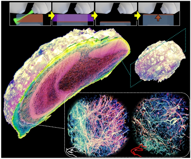
针对这一难题,研究组开发了空间精度可控的光化学切片技术。通过将连续光化学切片与晶格层光成像、PB级计算相结合,完成了对小鼠整颗嗅球中轴突与髓鞘结构的纳米级分辨率成像与三维重建。全嗅球范围内的有髓/无髓轴突分析,揭示了神经退行性大脑中轴突退行性变化与脱髓鞘/髓鞘形成障碍的独特模式,充分证明了该方法支撑PB至EB级超分辨研究的潜力。
附:英文原文
Title: Mesoscale volumetric fluorescence imaging at nanoscale resolution by photochemical sectioning
Author: Wei Wang, Xiongtao Ruan, Gaoxiang Liu, Daniel E. Milkie, Wenping Li, Eric Betzig, Srigokul Upadhyayula, Ruixuan Gao
Issue&Volume: 2025-10-16
Abstract: Optical nanoscopy of intact biological specimens has been transformed by recent advancements in hydrogel-based tissue clearing and expansion, enabling the imaging of cellular and subcellular structures with molecular contrast. However, existing high-resolution fluorescence microscopes are physically limited by objective-to-specimen distance, which prevents the study of whole-mount specimens without physical sectioning. To address this challenge, we developed a photochemical strategy for spatially precise sectioning of specimens. By combining serial photochemical sectioning with lattice light-sheet imaging and petabyte-scale computation, we imaged and reconstructed axons and myelin sheaths across entire mouse olfactory bulbs at nanoscale resolution. An olfactory bulb–wide analysis of myelinated and unmyelinated axons revealed distinctive patterns of axon degeneration and de-/dysmyelination in the neurodegenerative brain, highlighting the potential for peta- to exabyte-scale super-resolution studies using this approach.
DOI: adr9109
Source: https://www.science.org/doi/10.1126/science.adr9109